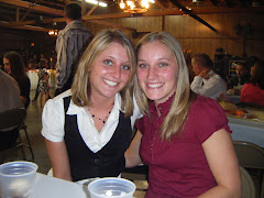
I choose this pathology because this happened to my mom about 18 years ago. She was playing 3rd base in a softball game, the batter hit a line drive right at her and before she had time to react the softball hit her right in the “cheek bone,” better known to us as the maxillary bone. This blunt trauma ended up fracturing part of her maxilla and zygoma. Today, she has 2 different metal plates in her face, one medially holding together her zygoma and one across her maxillary bone.
**What is a blowout fracture?**
A blowout fracture is a fracture to the orbital floor. Blowout fractures are caused by direct trauma to the globe that may cause decompression and pressure to the eye. This blunt trauma can be caused by things such as sports accidents, automobile accidents, or even occupational injuries. The orbits around the eye socket are very thin, with enough pressure they will break. When the bone breaks the globe and surrounding muscles tend to settle into the fracture. If this occurs the muscles can become trapped in the fracture and the injured eye will not move properly. Generally, the eye itself is not affected. Luckily for my mom, she did not experience any difficulties in the movement of her eye and has had no vision problems evolve since the accident.
**Symptoms**
Symptoms of a blowout fracture may include, but are not limited to, blurred vision, difficulty in moving the eye, swelling or bruising around the eye, double vision, blood in the white area of the eye, and eye pain. These symptoms usually take place right after the trauma occurs. In the instance of my mom, she had immediate swelling and bruising of the eye. There was blood in the white area of her eye and she experienced extreme pain when trying to move her eye. One other thing that she experienced was that her entire cheek was numb immediately after contact.
**Treatment**
In some cases, blowout fractures may heal themselves, but are carefully monitored. When surgery is not required it is mostly likely because a muscle is not trapped or less than 50% of the floor of the orbit was affected. In other cases, surgical repair is required. This is usually performed when an eye muscle is trapped in the fracture, or if there is extensive involvement of the bones of the orbital floor. Majority of surgeries to repair the orbital bones is done 1 to 2 weeks after the injury has occurred so that any swelling can subside. In most cases an implant is used for reconstruction. After surgical repair the patient’s eye sight must be monitored often until discharge. The patient should also be instructed to avoid strenuous activity and blowing their nose for at least 2 days. In the case of my mom, she was immediately taken to the hospital where x-rays were preformed. She was told by the doctor that there was no injury to her orbital area. Two weeks after the accident she still had a little swelling and bruising, but the pain had diminished to a tolerable level. She had a dentist appointment about 2 weeks after the accident, and as soon as her dentist evaluated her he suggested that she get another opinion because he believed that she had a fractured orbital floor. Two days after seeing her dentist and getting another doctor referral she was going to surgery to repair her zygoma and maxilla. The doctor placed 2 metallic rigid miniplates in her face.

Blowout fracture in a 14-year-old boy after a blow to the left eye. Coronal reformatted CT image obtained with soft-tissue window settings shows a thickened and rounded appearance of the inferior rectus muscle (*), a finding indicative of muscle impingement, and depicts a hemorrhage in the left maxillary sinus (arrowhead).

A classic radiographic finding in blow-out fractures is the presence of a polypoid mass (the tear-drop) protruding from the floor of the orbit into the maxillary antrum The tear-drop represents the herniated orbital contents, periorbital fat and inferior rectus muscle.

No comments:
Post a Comment